
Brain Scan for Anxiety: What the Science Actually Shows
August 2, 2026


No single brain scan can diagnose an anxiety disorder in an individual. That verdict comes directly from clinical guidelines and recent reviews, and it matters because many patients arrive at clinics hoping a scan will hand them a clear answer. What neuroimaging can do is meaningfully different: it reveals group-level patterns in brain activity and structure, rules out other neurological causes, and, when combined with psychosocial assessment, adds useful information to a clinical picture.
Here is what you will find in this guide:
The science of anxiety brain imaging has matured considerably, but it has also grown more honest about its limits. Across hundreds of studies, a few patterns show up consistently at the group level: the amygdala tends to be hyperreactive to threat cues, the anterior cingulate cortex (ACC) and insula show altered activation, and large-scale networks, including the default mode network (DMN), salience network, and central executive network, display abnormal connectivity in people with anxiety disorders compared to healthy controls.
A 2025 whole-brain resting-state fMRI study published in Frontiers in Psychiatry found that medication-naïve patients with generalized anxiety disorder (GAD) showed enhanced PCC–SMG connectivity that correlated strongly with Hamilton Anxiety Rating Scale scores (r = 0.667, p < 0.001). That is a meaningful correlation, but it describes a group average, not a reliable individual marker.
Statistic to know: A large-cohort analysis published in Translational Psychiatry examined a very large adult cohort and found that psychosocial variables, including depressive symptoms and childhood trauma history, dominated anxiety classification. Structural MRI features added only conditional improvements in specificity when combined with psychosocial data, not when used alone.
Why do findings vary so much across studies? Molecular Psychiatry (2024) points to heterogeneous network-level patterns across anxiety subtypes. GAD, panic disorder, social anxiety disorder, and PTSD each show partially distinct neural signatures, and even within a single diagnosis, individual variation is substantial. That heterogeneity is the core reason no single neural signature has been validated for individual-level diagnosis. Researchers in 2025–2026 describe a trajectory toward individualized “theranostic” biomarkers, but significant validation remains pending before any of those markers reach routine clinical use.
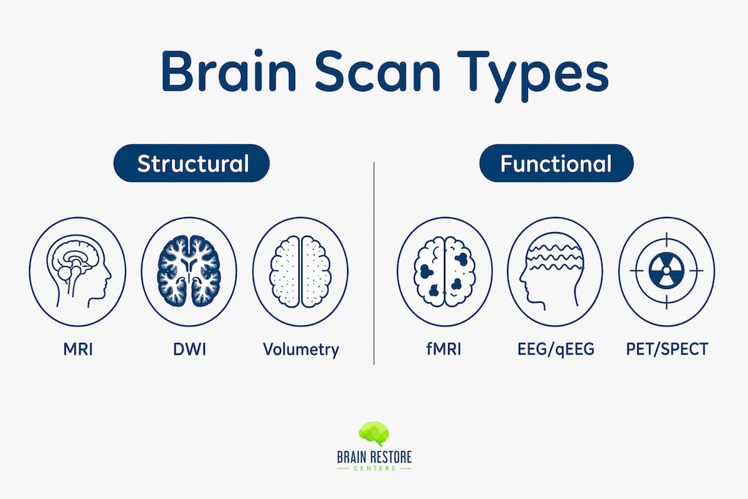
Understanding the differences between scan types helps you ask sharper questions when talking with a clinician. Each modality captures a different signal, carries different practical trade-offs, and serves different purposes in research versus clinical care.
Structural MRI maps the physical architecture of the brain: gray matter volume, white matter integrity, and regional size. It does not capture brain activity in real time. Clinically, it is most useful for ruling out structural causes of anxiety-like symptoms, such as tumors, lesions, or vascular changes. It is widely available, takes about half an hour to close to an hour, requires no radiation, and is covered by most insurance when there is a documented clinical indication. The main contraindications are metallic implants and severe claustrophobia.
Functional MRI (fMRI) measures the blood-oxygen-level-dependent (BOLD) signal, a proxy for neural activity. Task-based fMRI captures responses to specific stimuli (for example, viewing threatening images), while resting-state fMRI maps connectivity between brain regions when the patient is simply lying still. The Frontiers 2025 study cited above used resting-state fMRI and found connectivity changes that tracked with symptom severity in GAD. Resting-state fMRI is a promising treatment-response biomarker but remains largely a research tool. Clinical fMRI is available at academic medical centers and some large hospital systems; it is rarely covered by insurance for anxiety specifically.
Electroencephalography (EEG) records electrical activity across the scalp in real time. Quantitative EEG, or qEEG, applies statistical analysis to that data, comparing an individual’s brainwave patterns against a normative database. Clinics sometimes call this “brain mapping.” It is non-invasive, involves no radiation, and takes roughly about one to one and a half hours for a full assessment. qEEG is used clinically to guide neurofeedback protocols and, in some settings, to inform medication decisions. It does not diagnose anxiety disorders on its own, but it can identify dysregulated brainwave patterns that correlate with anxiety symptoms and help personalize treatment.
PET scans use a radioactive tracer to measure metabolic activity or receptor density in the brain. Early anxiety research used PET extensively to study glucose metabolism and neurotransmitter systems. Today, PET is primarily a research tool for anxiety; clinical use is largely limited to oncology, neurology, and dementia workups. It involves radiation exposure, requires IV tracer injection, and is expensive (typically $3,000–$6,000 or more without insurance). It is not a standard clinical option for anxiety evaluation.
SPECT measures regional cerebral blood flow using a radioactive tracer. Like PET, it involves radiation and is not a validated standalone diagnostic tool for anxiety. Some private clinics market SPECT-based “brain health assessments,” but no professional psychiatric or neurological guideline endorses SPECT as a diagnostic test for anxiety disorders. Approach such offerings with caution.
Comparison at a glance:
| Modality | What it measures | Clinical utility for anxiety | Availability | Procedure time | Radiation / risks |
|---|---|---|---|---|---|
| Structural MRI | Brain structure, volume | Rule-out neurological causes | Widely available | roughly half an hour to three quarters of an hour | None; claustrophobia risk |
| Resting-state fMRI | Functional connectivity (BOLD) | Research; emerging biomarker | Academic/specialist centers | approximately 45 minutes to over an hour | None; claustrophobia risk |
| EEG / qEEG | Electrical brain activity | Guides neurofeedback; symptom correlation | Specialty clinics | around one to one and a half hours | None |
| PET | Metabolism, receptor density | Research only | Research centers | around one to one and a half hours | Low-dose radiation; IV tracer |
| SPECT | Cerebral blood flow | Not guideline-endorsed for anxiety | Private clinics | half an hour to one hour | Low-dose radiation; IV tracer |
Pro Tip: Before agreeing to any scan, ask your provider one direct question: “What clinical decision will this result change?” If the answer is vague, the scan may not be worth the cost or the exposure.
Most anxiety diagnoses are made clinically, through structured interviews, validated symptom scales, and psychosocial history. Imaging enters the picture under specific circumstances.
Clinicians are likely to order imaging when:
Imaging is primarily research-only when:
The multimodal evidence is clear: psychosocial variables carry stronger predictive power for anxiety outcomes than neuroimaging alone. Imaging adds the most value when it is layered on top of a thorough clinical assessment, not substituted for one. For most patients with straightforward GAD or panic disorder, a brain scan will not change the treatment plan.
Pro Tip: When asking your provider about an imaging referral, try this framing: “Can you help me understand what finding on this scan would lead to a different treatment than what we’d do without it?” That question focuses the conversation on clinical utility rather than reassurance-seeking.
For guidance on how to frame these conversations with your care team, the resource on communicating mental health needs to your provider is a practical starting point.
qEEG brain mapping and neurofeedback occupy a distinct space between research-grade neuroimaging and conventional psychiatric care. They are clinic-facing services with a growing evidence base for symptom support, and understanding the patient pathway helps you evaluate any clinic offering them.
1. Intake and history review. The clinician collects a full symptom history, reviews any prior diagnoses or medications, and identifies treatment goals. This step shapes how the qEEG data will be interpreted.
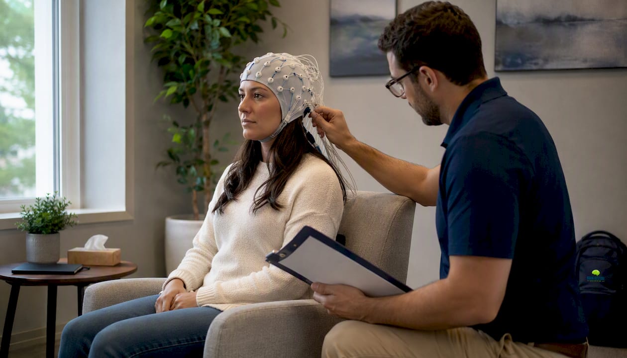
2. qEEG brain mapping session. A technician places an electrode cap on your scalp and records brainwave activity across multiple frequency bands (delta, theta, alpha, beta, gamma) for roughly 60–90 minutes. You sit quietly with eyes open and eyes closed during different recording segments. No needles, no radiation, no discomfort beyond the gel used to improve electrode contact.
3. Interpretation and protocol design. A licensed clinician with neurofeedback training compares your brainwave patterns against a normative database and identifies dysregulated areas. This informs which brain regions and frequency targets the neurofeedback protocol will address. You can learn more about what to expect from a qEEG brain mapping session at a clinic like Brainrestoremeridian.
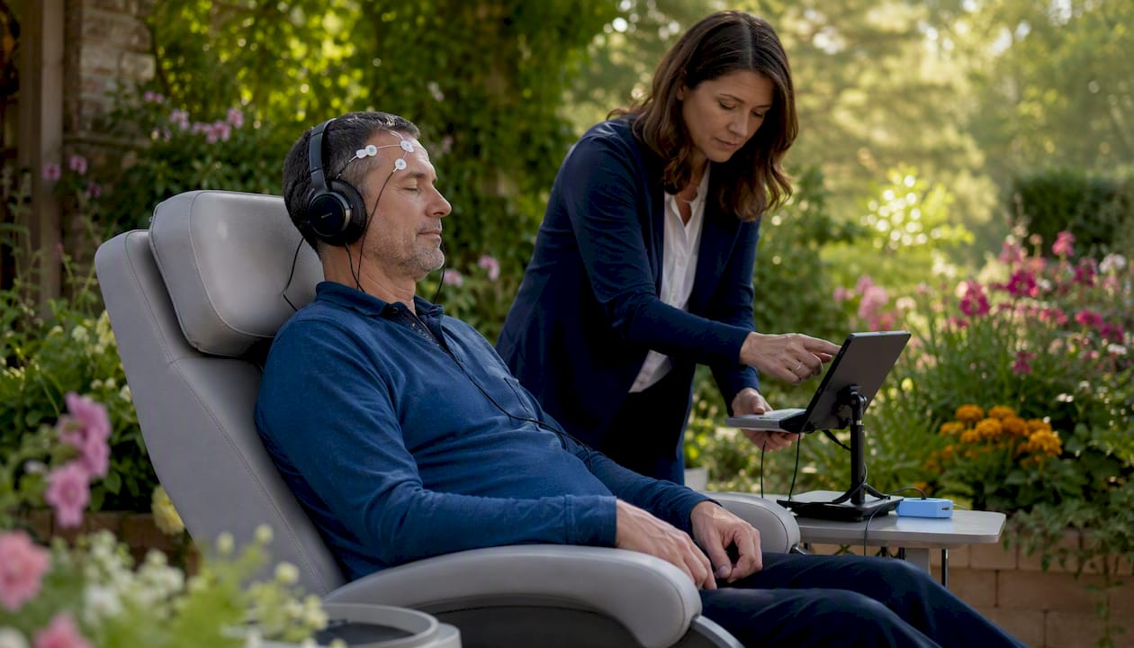
4. Neurofeedback trial sessions. Neurofeedback works by giving your brain real-time feedback, typically through audio or visual cues, when it produces target brainwave patterns. Think of it as guided practice for self-regulation. Sessions typically run 30–45 minutes, and a standard course ranges from 20–40 sessions depending on symptom severity and response.
5. Progress monitoring. A good clinic tracks outcomes with validated symptom scales at regular intervals, not just subjective patient report. Ask specifically how progress is measured and at what intervals.
Credentials to verify before committing to a provider:
Realistic expectations: Research supports neurofeedback for symptom reduction and improved self-regulation in anxiety. It is not a cure, and no reputable clinic should promise otherwise. The real benefits of neurofeedback for anxiety are meaningful but incremental, and they work best as part of a broader care plan.
Pro Tip: Ask three questions before starting: (1) How do you measure my progress, and with which validated scale? (2) How long do you retain my brain data, and who has access to it? (3) What is your protocol if I don’t respond after 10 sessions?
Brain scans are powerful tools, but they come with real limits and some genuine ethical concerns that deserve honest discussion.
Technical and interpretive limits:
Safety and contraindications:
Ethical and privacy concerns:
Red flags to watch for: Any clinic or vendor that claims a single scan diagnoses your anxiety disorder, guarantees specific symptom outcomes, or sells a proprietary scoring system without peer-reviewed validation is making claims the science does not support. The heterogeneity of anxiety’s neural signatures means no proprietary algorithm has been independently validated to reliably classify anxiety at the individual level. Walk away from any provider who tells you otherwise.
If you and your clinician decide imaging is appropriate, here is how to move through the process efficiently.
Steps to request imaging:
Approximate U.S. cost ranges:
Preparation and logistics:
For a practical overview of what local brain function testing involves, the brain function testing guide at Brainrestoremeridian walks through what patients can expect step by step.
Insurance notes: Medicare and most private insurers cover structural MRI when there is a documented neurological indication. Coverage for fMRI, qEEG, and neurofeedback varies significantly by plan. Call your insurer before scheduling and ask specifically whether the CPT code your provider plans to use is covered under your plan.
The research base for anxiety neuroimaging is large and uneven in quality. These are the sources worth your time.
Frontiers in Psychiatry, 2025 whole-brain rs-fMRI study: Best for understanding specific connectivity changes in medication-naïve GAD patients and how they correlate with symptom severity. A good starting point for clinicians interested in resting-state biomarkers.
Molecular Psychiatry, 2024 network-level review: The clearest current account of why a single neural signature for anxiety has not been validated. Useful for anyone trying to understand why neuroimaging findings vary so much across studies.
Translational Psychiatry / Nature, 2026 multimodal classification study: Essential reading for understanding the relative contribution of imaging versus psychosocial data. The NAKO cohort size (a very large adult cohort) makes this one of the most statistically powered studies available.
MedicalXpress summary, 2025: A patient-accessible summary of the current state of anxiety brain scanning. Good for sharing with family members or non-specialist providers.
Topics in Magnetic Resonance Imaging, 2024 guided imagery trial: Practical for clinicians and patients preparing for MRI. The evidence for guided imagery as a scan-anxiety intervention is solid.
Springer VR and preparatory techniques review, 2025: Covers video walkthroughs, guided imagery, and VR familiarization as tools to reduce MRI anxiety. Research-grade, clinic-applicable.
For readers interested in how qEEG fits into a broader brain health evaluation locally, Brainrestoremeridian’s comprehensive brain health restoration page describes how neuroimaging, neurofeedback, and functional medicine work together in an integrated care plan.
No brain scan currently available can diagnose an anxiety disorder on its own; imaging is most valuable when it is layered onto a thorough psychosocial assessment, not substituted for one.
| Point | Details |
|---|---|
| No standalone diagnostic scan | Clinical guidelines confirm no single brain scan reliably diagnoses anxiety at the individual level. |
| Group-level findings are real but limited | Amygdala hyperreactivity, ACC/insula changes, and network alterations are consistent group findings, not individual markers. |
| Psychosocial data dominates classification | A 26,378-adult study found psychosocial variables outperform structural MRI alone for classifying anxiety outcomes. |
| qEEG and neurofeedback are clinic-facing options | Both services have evidence for symptom support; verify provider credentials and outcome-tracking protocols before starting. |
| Brainrestoremeridian offers local evaluation | Brainrestoremeridian in Meridian, Idaho provides qEEG brain mapping and neurofeedback as part of an integrated, clinician-supervised care plan. |
The most common mistake I see in how brain scanning is discussed for anxiety is the framing of imaging as a verdict rather than a data point. Patients sometimes arrive hoping a scan will confirm what they already feel, and clinicians sometimes order imaging to satisfy that hope rather than to answer a specific clinical question. Neither serves the patient well.
What imaging does well, when ordered thoughtfully, is narrow the differential. A structural MRI that comes back clean is genuinely reassuring when a patient has new-onset anxiety after 45 with no prior psychiatric history. A qEEG that shows elevated high-beta activity in frontal regions gives a neurofeedback provider a starting point for protocol design. These are real contributions. But they are contributions to a clinical conversation, not substitutes for one.
The research trajectory is encouraging. The move toward multimodal models that combine imaging with psychosocial data is the right direction, and the 2025–2026 literature reflects that. For now, the most honest thing a clinician can say is: “This scan will help us rule things out and, in some cases, personalize your care. It will not hand us a diagnosis.” At Brainrestoremeridian, that framing guides every evaluation, from the initial intake through outcome tracking.
If you have been wondering whether a brain scan could help clarify what is driving your anxiety, Brainrestoremeridian in Meridian, Idaho offers a structured evaluation pathway built around that exact question. The clinic’s qEEG brain mapping service records your brainwave activity, compares it against a normative database, and produces an interpretive report that a licensed clinician reviews with you directly. From there, a personalized neurofeedback program for anxiety can be designed around your specific pattern, not a generic protocol.
Every evaluation at Brainrestoremeridian is presented as what it is: a complementary tool that informs care, not a standalone diagnosis. Outcome tracking with validated symptom scales is built into the process from the start. If you are ready to get a clearer picture of your brain’s activity and explore what targeted support might look like, you can book a qEEG evaluation or contact the clinic directly to ask questions before committing to anything.
This article is general health information, not medical advice. Please consult a qualified clinician for guidance specific to your situation.
Frontiers in Psychiatry, 2025 rs-fMRI study: Research-grade; best for clinicians and scientifically literate patients wanting to understand specific connectivity findings in GAD. The correlation data (r = 0.667 with Hamilton Anxiety scores) is among the strongest published for resting-state fMRI in this population.
Molecular Psychiatry, 2024 network review: High-authority review; explains why heterogeneous network patterns across anxiety subtypes prevent a single validated neural signature. Essential for understanding the limits of current neuroimaging.
Translational Psychiatry / Nature, 2026 multimodal study: Large-cohort, research-grade; the NAKO dataset (a very large adult cohort) makes this the most statistically robust multimodal classification study available. Useful for clinicians and researchers evaluating the incremental value of imaging.
MedicalXpress, 2025 summary: Patient-facing; accessible summary of the current state of anxiety brain scanning. Good for non-specialist audiences and for sharing with family members.
Topics in Magnetic Resonance Imaging, 2024 guided imagery trial: Clinic-applicable; the 88-patient randomized trial provides solid evidence for guided imagery as a practical intervention to reduce MRI-related anxiety. Useful for both patients and technologists.
Springer VR and preparatory techniques, 2025: Research-grade with clinic applications; covers video walkthroughs, guided imagery, and VR familiarization. Recommended for any clinic that performs MRI on anxious patients.
Questions about your next step?
Our Meridian team can help you understand the evaluation process and which care options may fit your needs.
Schedule a Brain Health ConsultationPrefer to speak with our team? Call 208-639-1397