Brain Scan for Memory Loss: Your Meridian Care Path
July 29, 2026

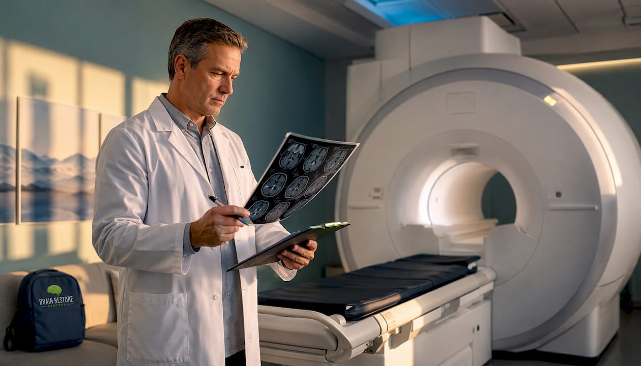
When memory concerns arise, a noncontrast MRI is typically the first brain scan ordered. It rules out structural causes like stroke, tumor, or fluid buildup before any other testing. Functional scans such as FDG-PET, SPECT, or amyloid PET come later, reserved for atypical presentations, early-onset cases (before age 60), or when MRI results leave the picture unclear. The American Academy of Family Physicians recommends specialist referral when symptoms begin before 60, behavioral changes are significant, or diagnosis remains uncertain after initial evaluation. As Mayo Clinic notes, early evaluation matters because it opens the door to treating reversible causes and planning care sooner.
Quick reference:
Structural and functional imaging answer different questions. Structural scans, MRI and CT, show the brain’s anatomy: atrophy patterns, stroke damage, tumors, vascular changes, and fluid accumulation. Functional scans, FDG-PET, SPECT, and amyloid PET, measure what the brain is doing: blood flow, glucose metabolism, and the presence of abnormal proteins like amyloid.
| Modality | Main use | What it shows | When ordered | Key constraints |
|---|---|---|---|---|
| MRI (noncontrast) | First-line structural workup | Atrophy, stroke, lesions, vascular disease, tumors | Initial evaluation | Metal implants, pacemakers; longer scan time |
| CT | Structural alternative | Hemorrhage, hydrocephalus, gross atrophy | MRI unavailable or contraindicated | Radiation exposure; less soft-tissue detail |
| FDG-PET | Metabolic activity | Glucose hypometabolism patterns | Atypical or unclear MRI; early-onset | Specialist order; prior auth often required |
| Amyloid PET | Protein deposition | Amyloid plaques (Alzheimer’s marker) | Unexplained MCI, targeted therapy consideration | High cost; not routine screening |
| SPECT | Blood flow | Regional perfusion patterns | MRI/CT inconclusive | Radiotracer injection; imaging |
| fMRI | Brain activity | Task-related blood flow changes | Primarily research; selected clinical cases | Limited clinical availability locally |
One point worth holding onto: a normal scan does not rule out early cognitive impairment. Imaging in early-stage disease can appear unremarkable, which is exactly why scans are interpreted alongside clinical exams, lab work, and cognitive testing, never in isolation.
“Many cases of memory loss are not progressive dementia. Distinguishing reversible causes through clinical evaluation is a primary goal.” — Merck Manual
A radiology report is one piece of a larger picture. Clinicians combine imaging with a cognitive screen such as the MoCA, lab panels (B12, TSH, metabolic panel), and a thorough medication review. The MoCA results can be influenced by factors such as education and cultural background, so when screening results and clinical impression conflict, formal neuropsychological testing offers higher diagnostic accuracy. Labs and medication review often surface reversible contributors, including thyroid dysfunction, vitamin deficiencies, or drug interactions, before expensive imaging is even needed.
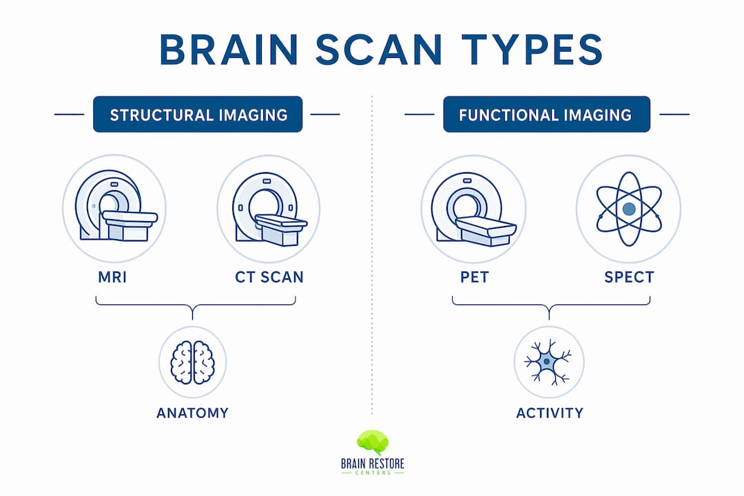
Functional scans like PET and SPECT are not routine. Most patients will not need them. They are ordered selectively for unexplained mild cognitive impairment, atypical presentations, or when a clinician is considering disease-specific therapy. If you are experiencing early onset of memory changes, or if symptoms are progressing quickly, that is a clear signal to pursue specialist evaluation rather than waiting.

Pro Tip: Before any appointment, update your medication and supplement list to include dosages and how long you have been taking each one. Clinicians review this before ordering imaging because many common drugs and supplements can cause reversible cognitive symptoms that look like early dementia on screening.
Local availability matters too. MRI and CT are widely accessible in the Meridian and greater Boise area. Amyloid PET and advanced fMRI typically require referral to a regional imaging center. Sleep health is also worth factoring in: poor sleep may be linked to increased risk of Alzheimer’s disease, and it is a modifiable factor your care team can address alongside imaging. After imaging, physical therapy for neurological conditions is often part of a recovery plan, particularly when vascular or structural findings are present.
Waiting on referrals and piecing together imaging reports, lab results, and specialist notes on your own is exhausting. Brainrestoremeridian, operating as Align Integrated Medical Clinic in Meridian, Idaho, offers a different starting point: a coordinated intake that reviews your prior scans, orders MRI or CT locally when needed, arranges referrals for PET or specialized imaging, and builds a personalized care plan around what the results actually show.
The clinic’s brain health restoration program combines neurofeedback, photobiomodulation (laser therapy), hyperbaric oxygen therapy, functional-medicine lab work, chiropractic integration, and progressive rehabilitation. These are not add-ons. They are built into the care plan from the start, alongside imaging coordination and specialist referrals when indicated. The clinic also integrates formal neuropsychological testing when cognitive screening results need a deeper look. If you are ready to move from uncertainty to a clear next step, schedule an intake at Brainrestoremeridian and bring your medication list and any prior imaging reports.
A noncontrast MRI is the standard first scan for memory loss evaluation, with functional imaging reserved for atypical, early-onset, or diagnostically unclear cases.
| Point | Details |
|---|---|
| First-line scan | Noncontrast MRI rules out structural causes; CT is the alternative when MRI is unavailable. |
| Functional imaging is selective | FDG-PET, amyloid PET, and SPECT are ordered for atypical, early-onset, or unclear cases, not routine screening. |
| Normal scan ≠ no problem | A normal scan does not exclude early cognitive impairment; imaging is always interpreted with labs and clinical testing. |
| Medication review comes first | A complete medication and supplement list can uncover reversible causes before imaging is ordered. |
| Brainrestoremeridian | Coordinates local imaging, specialist referrals, and restorative therapies including neurofeedback and hyperbaric oxygen in Meridian, Idaho. |
This article is general health information, not medical advice. Confirm your specific situation with a qualified clinician or primary care provider.